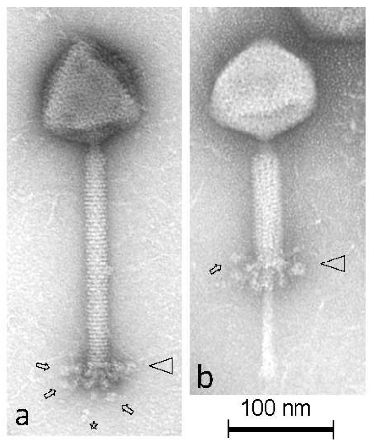
Figure 1.
Transmission electron micrographs of L. plantarum phage Dionysus negatively stained with 2% (w/v) uranyl acetate. Triangles and arrows indicate the terminal baseplate structure and representative flexible appendages with terminal globular structures, respectively, attached underneath them. The single distal tail fibers terminating with three distinct knob-like structures are indicated by the star symbol. Phage Dionysus is shown with extended tail sheath (a) and contracted tail sheath (b), substantiating that it is a Myoviridae phage.

