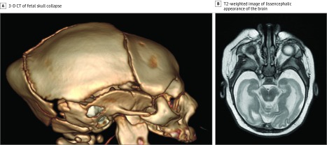
Figure 2. Neuroimaging Findings of an Infant With Severe Zika Virus.
A, Three-dimensional (3-D) computed tomography (CT) reconstruction demonstrates classic phenotypic pattern of fetal skull collapse with overlapping cranial sutures and prominent occipital protrusion. B, T2-weighted imaging demonstrates simplified gyral pattern with a lissencephalic appearance of the brain.

