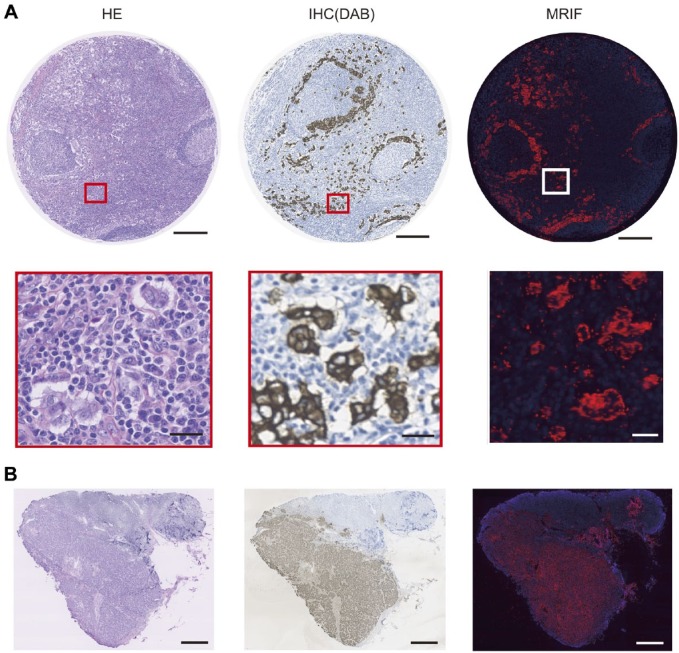
Figure 6.
Staining image of human breast cancer metastatic lymph node by HE, IHC (DAB), and MRIF treated with anti-pan-cytokeratin antibody-coated FF beads. (A) Image of a paraffin-embedded tissue array of human breast cancer metastatic lymph node stained with HE, IHC (DAB), and MRIF. Scale bar = 250 μm and 25 μm for high magnification. (B) Image of frozen tissue section of human breast cancer metastatic lymph node stained with HE, IHC (DAB), and MRIF. Scale bar = 1000 μm. Abbreviations: HE, hematoxylin and eosin; DAB, diaminobenzidine; MRIF, magnetically promoted rapid immunofluorescence; FF, fluorescent ferrite.

