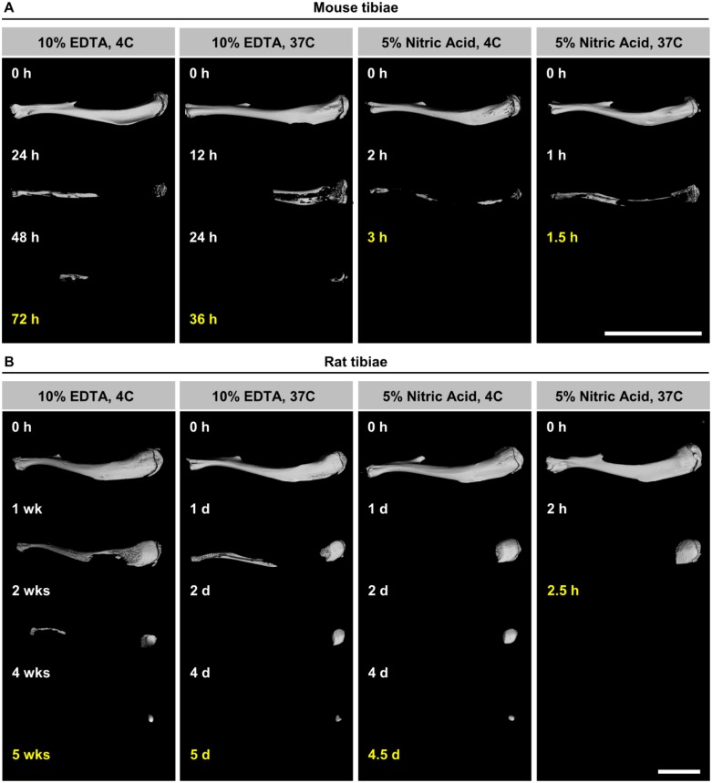
Figure 2.
Representative 3D microCT images of mouse (A) and rat (B) tibiae decalcified in 10% EDTA (slowest decalcification) and 5% nitric acid (fastest decalcification) at indicated time points with end points in yellow. Mineralized bone matrix appears white and decalcified bone is not visible. Scale bar = 1 cm. Abbreviations: 3D, three-dimensional; EDTA, ethylenediaminetetraacetic acid; microCT, micro–computed tomography.

