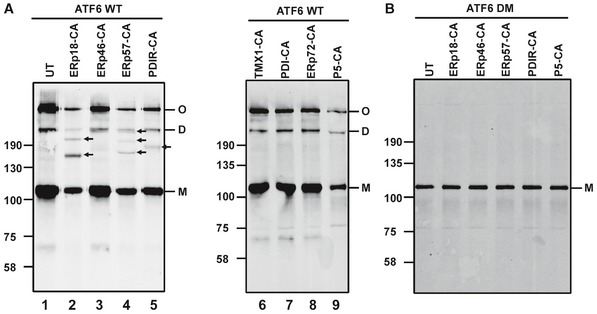
Figure 2. PDI family substrate‐trapping mutants form mixed‐disulfide complexes with ATF6.

- HEK293T cells stably expressing ATF6α were either left untransfected (UT) or transfected with substrate‐trapping mutants of PDI oxidoreductases. ATF6α was immunoisolated from cell lysates with mouse anti‐ATF6α and separated by SDS–PAGE under non‐reducing conditions and ATF6α detected by Western blotting using rabbit anti‐HA. Blots confirm mixed‐disulfide complexes between ATF6α and ERp18, ERp57, and PDIR indicated with arrows (lanes 2, 4, and 5). M, D, and O refer to ATF6α monomer, dimer, and oligomer, respectively.
- Whole‐cell lysates of HEK293T cells expressing an ATF6α mutant containing cysteine‐to‐alanine mutations in the lumenal domain (ATF6 DM) were transfected with PDI substrate‐trapping mutants. ATF6α was immunoisolated with mouse anti‐ATF6α and separated by SDS–PAGE under non‐reducing conditions and ATF6α detected with rabbit anti‐HA.
Source data are available online for this figure.
