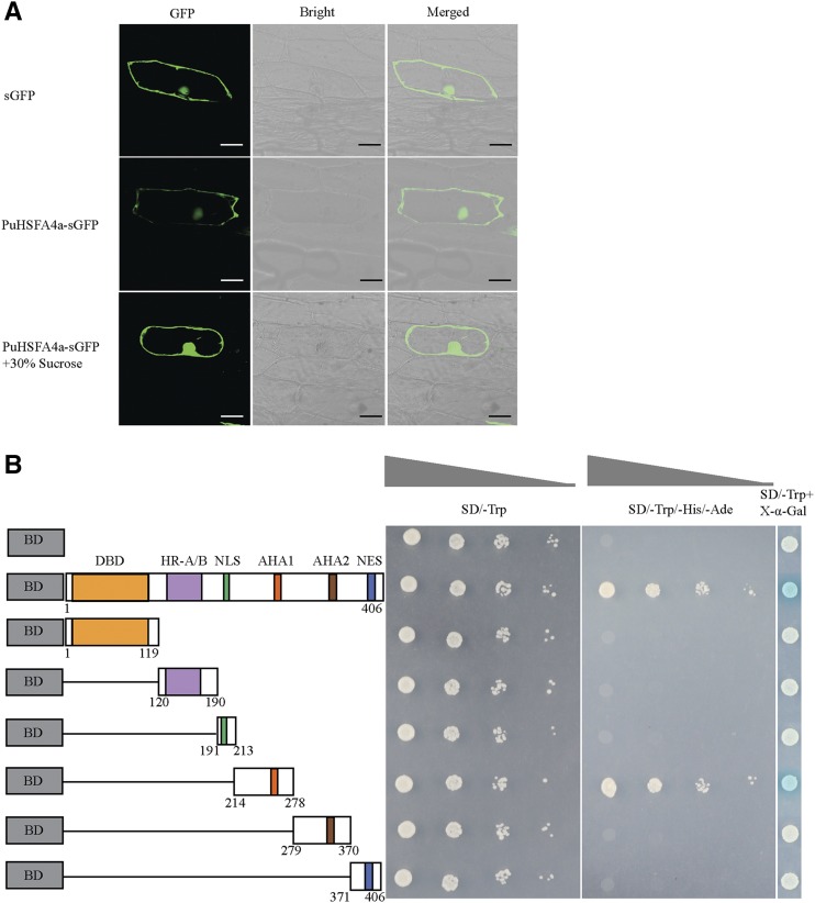
Figure 3.
Subcellular localization and transactivation assay of PuHSFA4a. A, Subcellular localization of GFP fusion PuHSFA4a protein in onion epidermal cells. Images were obtained in a dark field to detect green fluorescence, and again in bright light to observe the morphological characteristics of the cells. Bars = 50 μm. B, Transactivation assay of PuHSFA4a in yeast cells. The full-length construct and several partial deletion constructs of PuHSFA4a were fused to GAL4 DBD and expressed in the yeast strain AH109 Gold. Transformed yeast was grown in either SD/-Trp or SD/-Trp/-His/-Ade media. pGBKT7 was used as a negative control. LacZ activity was observed in SD/-Trp medium containing X-α-Gal.

