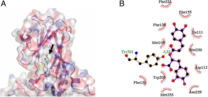
Fig. 3.
Molecular docking of rosmarinic acid (RA) to the VP1 of EV71. a Transparent view of the structure of RA (green) docked in the hydrophobic pocket within VP1 (purple). b Two-dimensional representation of hydrophobic interactions and hydrogen bonding within the VP1–RA complex. The side chains of residues forming the hydrophobic interaction are shown as arcs. A hydrogen bond was predicted in the Tyr201 of VP1, as shown by the dashed line. Location of RA is indicated by an arrow

