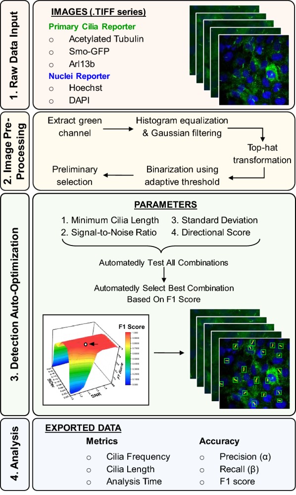
Fig. 1.

Overview flow diagram of Automated Cilia Detection in Cells (ACDC) software. The ACDC software for automatic detection of cilia comprises four main steps, which are lightly color-coded in four boxes: data input (red), pre-processing (yellow), detection (green), and analysis (blue); later figures follow this color-coding scheme. Step One: users can import hundreds of microscope images that comprise a cilia reporter and a nuclei reporter. Step Two: the images are pre-processed to facilitate formation of a binary mask. Step Three: based on a manually marked-up representative image, the software auto-optimizes the detection of true candidates by determining which combination of the four listed parameters yields the greatest accuracy score (F1 score). The parameter combination is then used to automatically detect true candidates from images of the same or similar image series. Step Four: analysis and exportation of data, which includes accuracy metrics such as false-positive (FP) and false-negative (FN) rates and typical cilia metrics such as frequency and length. The analysis time is also reported for comparison between manual, automated, and semi-automated analysis (i.e., automated analysis mode with the ability to manually correct detection errors)
