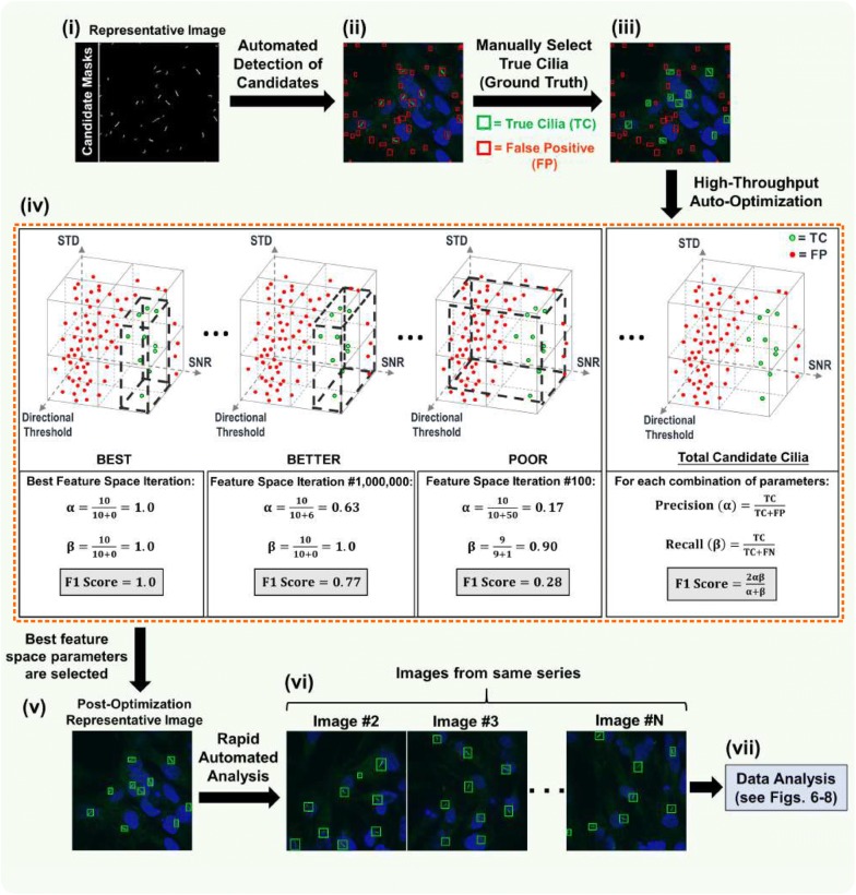
Fig. 4.
Automated detection optimization of candidate cilia. Auto-optimization of cilia detection consists of seven steps (i–vii). (i) The resulting image of the pre-processing step. Because this was the first image of the imported data series, it is used as a representative image for training and calibration. (ii) Initially, all post-binarization candidates considered to be false objects (outlined in red). (iii) Users must tell the software which candidates to accept by clicking on those true candidate cilia (now outlined in green). (iv) After the “Auto Parameters” feature is selected (see Fig. 3b; orange dashed box), the remainder of the analysis process is automated. Based on the candidates that were manually selected to be true, the software sifts through ~ 5.5 million iterations of parameter combinations and chooses the feature space (depicted as dashed black boxes) that encloses only the true candidates and excludes the other false candidates. The software determines the accuracy of each iterative trial by searching for the feature space that yields that greatest F1 score. The F1 score is a function of two variables—precision (α), which describes the frequency of false positives (FPs), and recall (β), which describes the frequency of false negatives (FNs). Here, the automatically detected true candidates are represented by ‘TC.’ (v–vi) This best feature space is used to automatically analyze the remainder of the image series. If necessary, the user has the ability to adjust the optimized feature space to their preference. (vii) After analysis of the last image in the series, the data are exported for further study

