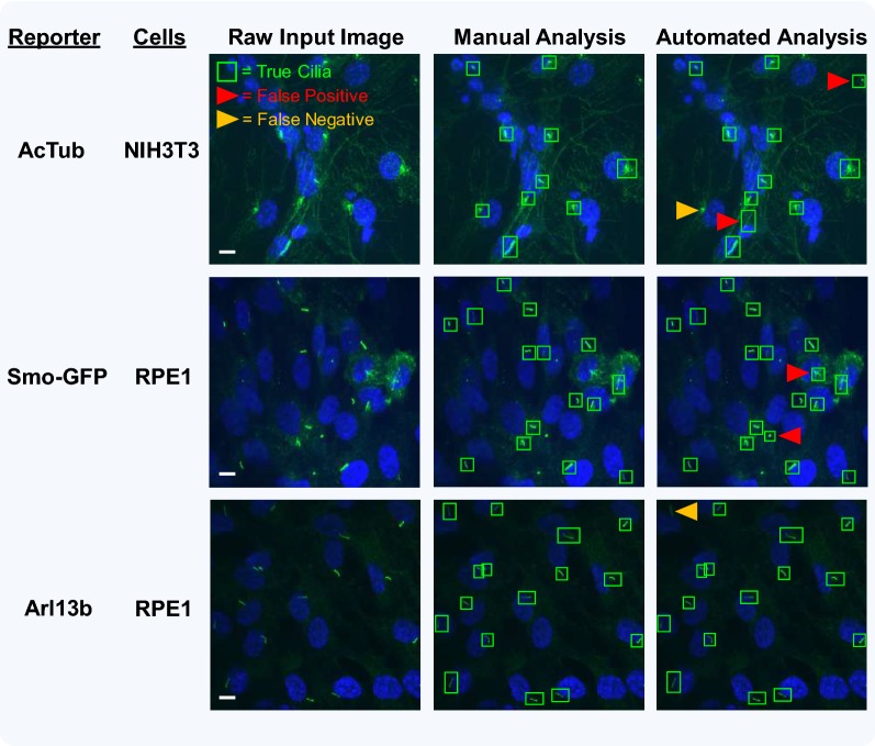
Fig. 5.
Identification of false-positive and false-negative candidate cilia during automated analysis with ACDC software. In order to evaluate how the rates of false positives (FPs) and false negatives (FNs) change across conditions, two different cell types (NIH3T3 and htert-RPE1) and three different ciliary reporters (AcTub, Smo-GFP, Arl13b) were analyzed. Left column: examples of raw input images. Middle column: the software’s “full manual analysis” mode was used to detect and accept every candidate based on user criteria. Right column: the software’s “automated analysis” mode was used to detect cilia. As shown, most of the manually selected true cilia were detected. However, there were occasionally some FPs (red arrowheads) and FNs (orange arrowheads). Note: for these representative images, FPs and FNs were identified manually (i.e., during semi-automated analysis). Images were acquired with a ×60 (1.4 NA) magnification objective. Scale bars = 10 μm

