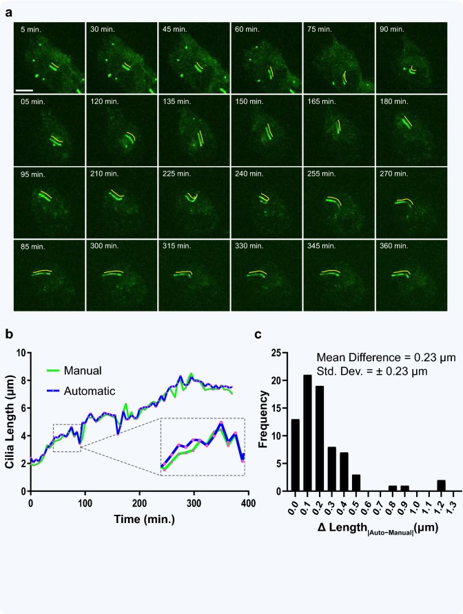
Fig. 8.
Application of ACDC to automated analysis of cilia movies. a A gallery of images from a live-cell spinning-disk confocal microscopy movie (370 min, 60× magnification) of a single cilium from an htert-RPE1 cell stably expressing Smoothened-pHluorin GFP, serum-starved and treated with 100 nM CytoD to promote ciliogenesis. Image frames of the GFP channel were taken from every 5 min and analyzed with the ACDC software. Yellow lines (shifted 10 pixels up and 10 pixels right) show the cilia ‘spline’ generated by the software during automated length measurement. Scale bar = 5 μm. b Movie frames were both manually and automatically analyzed for cilia length (0.143 μm/pixel). Line graph shows cilium growth over time; pink data points on both lines represent the analyzed frames. c Histogram of length measurement differences between manual and automated analysis reveals only a nominal difference of less than 2 pixels

