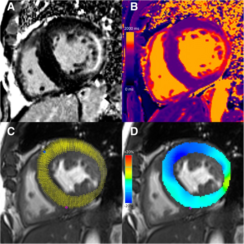
Fig. 1.

Cardiovascular magnetic resonance images of a 57-year-old male with Fabry disease. Short-axis late gadolinium enhanced (LGE) image demonstrates concentric left ventricular hypertrophy and mid-wall LGE at the inferior lateral wall (a). Short-axis native T1 map (native T1 value 1100 ms at the interventricular septum) (b). Short-axis cine balanced steady state free precession (bSSFP) image with circumferential myocardial strain analysis points (c) and color myocardial circumferential strain map (d). Global longitudinal strain was − 10.4%. Global circumferential strain was − 15.0%. Base-to-apex longitudinal strain gradient was 2.6%. Base-to-apex circumferential strain gradient was 2.1%
