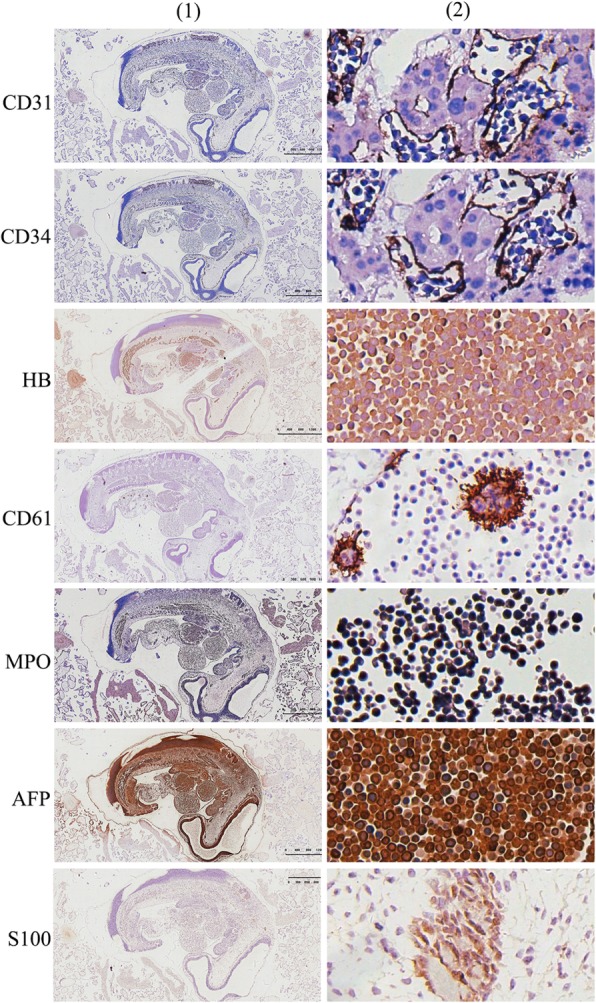
Fig. 2.

Immunohistochemical positive staining in the early embryonic tissue and its extramedullary hematopoietic tissue. CD31 positive staining in the vascular endothelial cells, suggesting that all the extramedullary hematopoietic cells were located in the blood vessels or naive liver sinus; CD34 positive staining in the vascular endothelial cells, suggesting that all the extramedullary hematopoietic cells were located in the blood vessels or naive liver sinus; CD61 positive staining in megakaryocytic cells, confirming that the positive cells are megakaryocyte; MPO positive staining in extramedullary hematopoietic celsl in the blood vessels, confirming that the positive cells are medullary hematopoietic cells; AFP positive staining in the extramedullary hematopoietic cells; S-100 positive staining in the neural tube; (1): 10× magnification; (2): 200× magnification)
