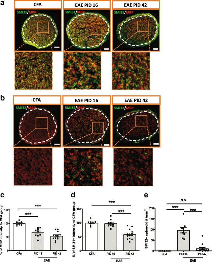
Fig. 5.
Demyelination and axonal injury were evident at early stage of EAE while axonal loss only was seen at the late stage of EAE. a SMI31 and MBP staining in the optic nerve of EAE mice. b SMI32 and MBP staining in the optic nerve of EAE mice. c-d Quantification of MBP and SMI31 as in figure a. e Quantification of SMI32 spheroid staining as in figure b. Boxed regions from each images were magnified as shown in figures. White dash line delineated ROI. Quantification is shown as mean ± SEM. Significance was determined by two-tailed, unpaired Student’s t-test with P < 0.05 was considered as significant. *** P ≤ 0.001. N.S. = no significant difference. Scale bar =50 μm

