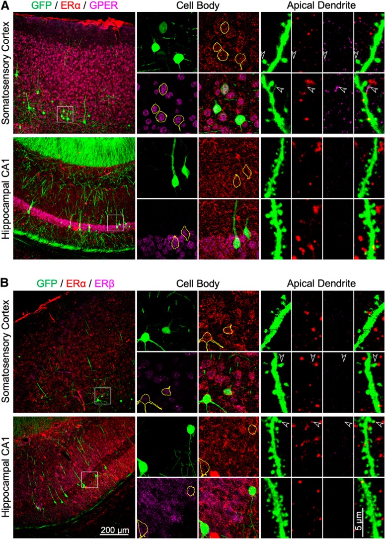
Figure 4.
Immunohistochemical staining of ERα, ERβ, and GPER in the somatosensory cortex and hippocampal CA1 in intact female Thy1M-GFP mice. A, Representative images showing immunostaining of GFP (green), ERα (red), and GPER (magenta) in the soma and distal apical dendrite of neocortical layer 5 and CA1 neurons. GFP-positive somata have been outlined in yellow. The arrowheads indicate colocalization of immunoreactivity in some dendritic spines or filopodia. B, Representative images showing the immunostaining of GFP (green), ERα (red), and ERβ (magenta) in the soma and apical dendrite of layer 5 and CA1 neurons.

