
Figure 2.
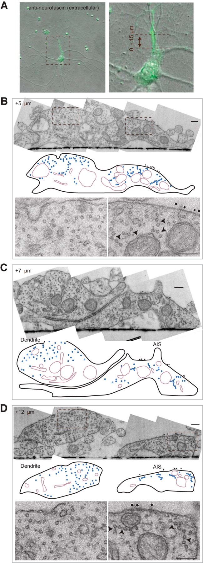
21 DIV neurons have fasciculated microtubules in the AIS. A–D, Typical example of a 21 DIV neuron processed by our correlative approach to analyze the AIS microtubules. Extracellular neurofascin staining to identify the AIS by fluorescence (A) and by EM (B–D). Note that there is a dendrite running parallel to the AIS, which at +5 μm did not split yet from the axon although the neurofascin marker and fasciculated microtubules already group at the right side of the neurite (B). In the schematic representations the microtubules and cross-bridges are represented in blue and organelles in violet. In the AIS magnifications arrowheads point to electron dense cross-bridges. Scale bar, 200 nm.