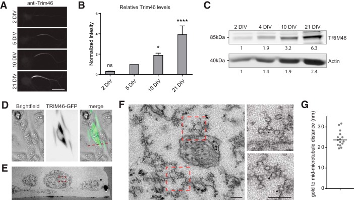
Figure 5.
TRIM46 localizes to the electron dense cross-bridge. A, Immunolabeling against TRIM46 at the AIS at different developmental stages. Scale bar, 20 μm. B, Quantification of TRIM46 labeling during neuron development, normalized to 5 DIV. N > 3 experiments of min 10 neurons each. Error bars represent SEM. Statistics, one-way ANOVA with Dunn's correction (compared with 5 DIV). ns, Nonsignificant; *p < 0.05, ****p < 0.0001. C, Western blot analysis of hippocampal neurons at different developmental stages. Neurons were grown at 500,000/well, lysed in 100 μl lysis buffer and equal amounts of cell lysate were loaded and stained for TRIM46. D, GFP-TRIM46 expression in HeLa cells. E, EM image of the transfected HeLa cell in D (as marked by the red dotted line). F, G, High-magnification of the boxed area in E, with further zooms showing the fasciculated microtubules upon GFP-TRIM46 overexpression in HeLa cells with immunolabeling with the gold-conjugated anti-gfp nanobody (F). G, The quantification of the gold to mid-microtubule distance and the black lines represent the median value. Scale bar, 200 nm.

