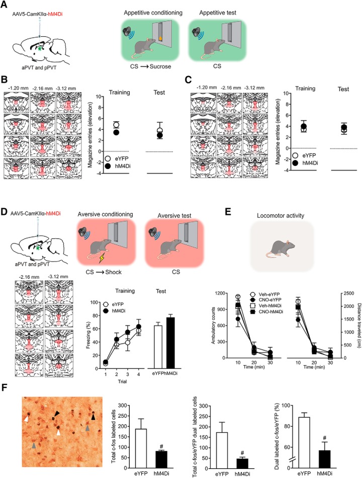
Figure 4.
A, Experiments 5a and 5b. AAV was used to express eYFP or hM4Di in PVT and rats then received appetitive conditioning using liquid (Experiment 5a) or pellet (Experiment 5b) reward. B, Experiment 5a. Placement map showing hM4Di expression with each animal represented at 10% opacity, eYFP (n = 8) or hM4Di (n = 8). Mean ± SEM appetitive responses at the end of appetitive conditioning and during test. Chemogenetic silencing of PVT had no effect on appetitive behaviors. C, Experiment 5b. Placement map showing hM4Di expression with each animal represented at 10% opacity in PVT, eYFP (n = 8) or hM4Di (n = 7). Mean ± SEM appetitive responses at the end of appetitive conditioning and during test. Chemogenetic silencing of PVT on test had no effect on appetitive behaviors. D, Experiment 5c. AAV was used express eYFP (n = 8) or hM4Di (n = 8) in PVT and rats then received aversive conditioning. Placement map showing hM4Di expression with each animal represented at 10% opacity. Mean ± SEM aversive responses during aversive conditioning and during test. Chemogenetic silencing of PVT had no effect on aversive behaviors. E, Experiment 5d. Chemogenetic silencing of PVT also had no effect on locomotor activity when assessed in an open field. F, Experiment 6. c-Fos immunohistochemistry was used to verify PVT chemogenetic inhibition, group hM4Di n = 8, eYFP n = 8. Example of single Fos (black arrowhead), single eYFP (gray arrowhead), and dual-labeled c-Fos/eYFP neurons (white arrowhead) in PVT. Mean ± SEM numbers of total c-Fos, total dual-labeled c-Fos/eYFP, and percentage dual-labeled c-Fos/eYFP neurons in PVT after CNO injection. #p < 0.05.

