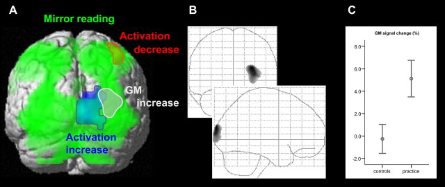
Figure 2.
Practice-related changes in activation and gray matter. A, Clusters of activation and GM changes are displayed at a height threshold of 0.001 uncorrected and an extent threshold of 0.05 corrected. Mirror-reading-specific activation (green), practice-related decrease of activation (red), practice-related increase of activation (blue), and practice-related increase of GM (gray) are superimposed on a surface-rendered MNI template. Compared with normal reading, mirror reading resulted in a differential activation of bilateral dorsal occipital lobe (inferior, middle, and superior occipital gyrus), occipitotemporal cortex (fusiform gyrus), superior parietal cortex (bilateral superior parietal lobule, left intraparietal sulcus, bilateral precuneus, and left somatosensory cortex), medial and dorsolateral prefrontal cortex (left presupplementary motor area, right middle cingulate cortex, left frontal eye field, and bilateral precentral gyrus), right anterior insula, and cerebellum. The comparison of GM before and after practice shows a significant increase in GM in a subset of the regions (gray) activated in the right occipital cortex during mirror reading (green). B, The middle plot shows the maximum intensity projection of the GM increase. C, The right plot displays the GM signal change (as a percentage) of all voxels of the cluster (we divided the individual mean of the GM values of all voxels of the resulting cluster after practice by the individual mean of the GM values of the same voxels before practice; error bars indicate mean and SEM). The mean relative GM signal increase within the reported cluster was 5.1% (SEM, 1.6%) in the practice group and −0.3% in the control group (SEM, 1.3%).

