
Figure 3.
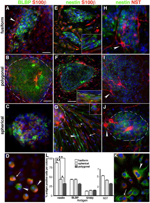
Cells in E13.5 OE colony cores and their progeny express neuronal, glial, and radial glial antigens. A–C, Fusiform, polygonal, and spherical colony cores contain cells expressing BLBP (green) and/or S100β (red). D, BLBP and S100β are asymmetrically expressed in dividing (arrowheads) and nondividing (arrow) spherical colony progeny cells. E–J, Nestin (green) is expressed in the cores of each colony subtype, and S100β (red) expressing cells (E–G) are outside colony cores and clustered at their edges (E, F, insets). G, Nestin+ cells with radial glia-like morphology (arrowhead) radiate out from a spherical colony, and are distinct from either nestin+/S100β+ (asterisk) or nestin−/S100β+ (arrow) cells with glial morphology. Dividing cells segregate nestin and S100β to individual daughter cells (G, inset). H–J, NST+ (red) nestin-negative neurons (arrowheads) are atop nestin+ cells found in all colony subtype cores. K, A dividing colony progeny cell coexpresses nestin and perinuclear NST (arrowhead), which can be distributed to individual daughter cells after division (arrow). L, The percentage of nestin, BLBP, S100β, or NST-positive cells in cores of individual colony subtypes cultured in EGF plus FGF for 10 DIV. There are significantly more nestin+ cells in fusiform colony cores compared with spherical or polygonal cores, and significantly more in spherical cores than polygonal cores (**p < 0.001, *p < 0.01; n = 3, average of 33 colonies tested per antigen). Error bars indicate SEM. The blue nuclear stain is DAPI. The dotted line indicates the edge of colony core. Scale bars: (in A) A, C–E, G, H, J, K, insets, 25 μm; (in B) B, I, 50 μm; F, 100 μm.