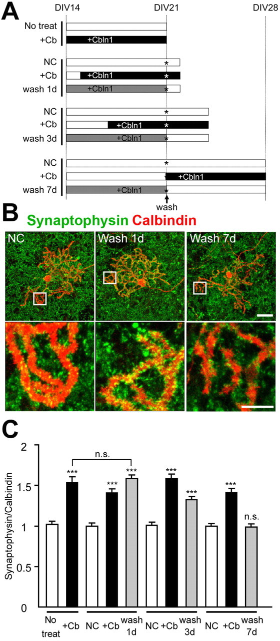
Figure 2.

Continued presence of Cbln1 is necessary to maintain cbln1-null Purkinje cell synapses in vitro. A, Experimental design. Cbln1-null cerebellar cultures were incubated with exogenous HA-Cbln1 (3 μg/ml) for 7 d from 14 DIV (gray bars). The cells were fixed on days 1, 3, or 7 after the removal of HA-Cbln1 at 21 DIV (wash 1 d, wash 3 d, and wash 7 d). As a negative control, the cells were incubated with medium containing no HA-Cbln1 for 7 d (No treat) or for indicated durations (NC, negative control; unfilled bars). As a positive control, HA-Cbln1 was added (3 μg/ml) to the medium at various time points (+Cb; filled bars). The culture medium was replaced and rinsed with the appropriate medium at 21 DIV (the asterisk indicates wash). B, Representative images of distal dendrites of Purkinje cells stained with calbindin (red) and synaptophysin (green). The synaptophysin signal on the dendrites gradually decreased. Scale bars: top, 30 μm; bottom, 10 μm. C, Average intensity of synaptophysin signal at the distal dendrites of the Purkinje cells. The effect of removing HA-Cbln1 was estimated by comparing with the no-treatment control (No treat) or negative control (NC) for each group. One day after the removal of exogenous Cbln1, the signal was still comparable with the level before removal, whereas 7 d after the removal, the signal declined to the value of the negative control (n = 24 cells; 3 independent experiments; ***p < 0.001). n.s., No significant difference.
