Figure 5.
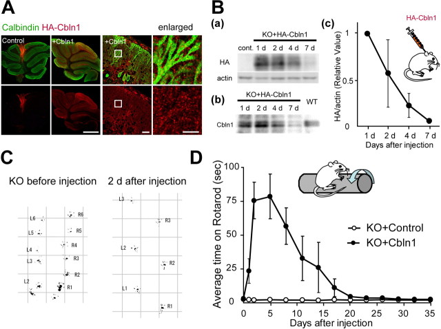
Improvement of cerebellar ataxia in adult cbln1-null mice after the subarachnoidal injection of Cbln1. A, Distribution of injected Cbln1, stained with anti-HA antibody (red), in the cerebellum at 24 h after injection. Purkinje cells were stained with calbindin (green). The injected Cbln1 was localized in all the cerebellar lobules. Enlarged images show the punctate signal of HA-Cbln1 localized on the dendrites of Purkinje cells. Scale bars: two far left columns, 1 mm; third column from the left, 30 μm; fourth column, 10 μm. B, Injected Cbln1 from the P2 fraction of the whole cerebellum was detected using immunoblotting and an antibody against HA (a) or Cbln1 (b) at 1, 2, 4, and 7 d after the injection. The signal in a was normalized with that of actin and further normalized to the value at 1 d after the injection (c). The injected HA-Cbln1 level decreased to <10% on day 7 (n = 3 mice). C, Representative gait pattern of a cbln1-null mouse before and 2 d after the injection. The hindpaws were marked with black paint. The irregular, shortened gait skips were markedly improved after the injection of Cbln1. L, Left paw; R, right paw. Numbers indicate step counts. D, Results on the rotarod test after the injection of Cbln1 medium (KO+Cbln1) or the control medium (KO+control). The average time on the rotarod at 20 rpm was calculated from four trials (n = 4 mice).

