Figure 7.

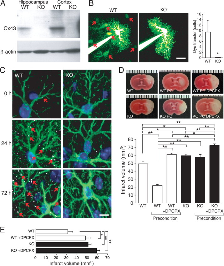
Cx43 expression is required for preconditioning in vivo. A, Western blot of hippocampus and cortex from a WT mouse (Cx43fl/fl) and from a Cx43 KO mouse (Cx43fl/fl:hGFAP–Cre). B, Intercellular diffusion of Lucifer yellow in astrocytes in slices from WT (red arrows indicate Lucifer yellow labeling of neighboring cells) and KO mice. Hippocampal slices were prepared from P18–P25 mice. Histogram compares diffusion of Lucifer yellow into neighboring cells in WT and KO. Only Lucifer yellow-positive cells within the field of view was included in the analysis (150 × 150 μm). Scale bar, 30 μm. Data represent average ± SE (n = 4–8). *p < 0.05, Tukey–Kramer test. C, Cx43 (white, small red arrows) and GFAP (green) immunohistochemistry from wild-type and Cx43 KO before or 24 and 72 h after preconditioning. Nuclei were stained with DAPI (blue). D, Comparison of infarct volume after MCA occlusion, preconditioning and MCA occlusion, and preconditioned mice that received one injection of the adenosine A1 receptors antagonist DPCPX (1 mg/kg, i.p.) immediately before hypoxic preconditioning. Data represent average ± SE (n = 6–8). E, Comparison of infarct volumes in mice with 30 min occlusion of the MCA. DPCPX-treated mice received an injection of 1 mg/kg intraperitoneally immediately before the occlusion. Data represent average ± SE (n = 6–8). *p < 0.05, **p < 0.01, Tukey–Kramer test.
