
Figure 2.
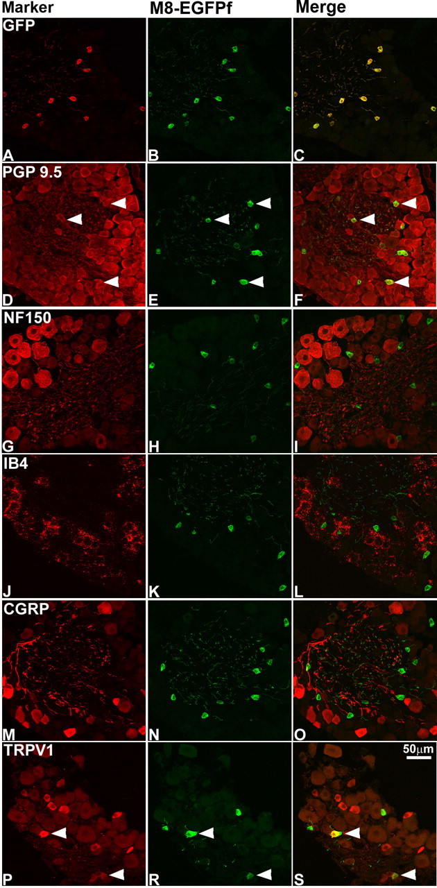
TRPM8EGFPf marks a unique population of DRG cell bodies. A–S, DRG neurons from TRPM8EGFPf/EGFPf mice were stained with antibodies against GFP and sensory neuron markers (red). TRPM8EGFPf neurons were visualized by endogenous EGFPf (green). Arrowheads indicate examples of double-labeled neurons. Scale bar (in S) is the same for all panels.