
Figure 4.
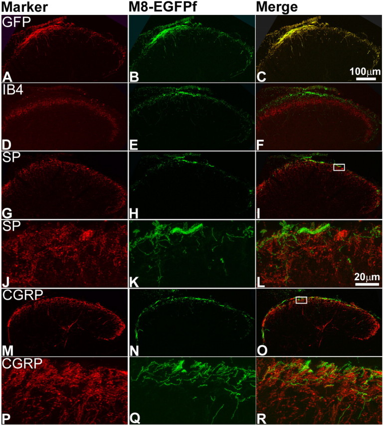
TRPM8EGFPf neurons terminate in lamina I of the spinal cord dorsal horn. A–R, Confocal images of the lumbar region of adult spinal cord from TRPM8EGFPf/EGFPf mice were stained with antibodies against GFP or sensory neuron markers (red). TRPM8EGFPf neurons were visualized by endogenous EGFPf (green). The boxed regions in I and O were reimaged at 60× with 2× digital zoom and shown in J–L and P–R, respectively. SP, Substance P. Scale bar (in C) is the same for A–I, M–O. Scale bar (in L) is the same for J–L, P–R.