Figure 2.

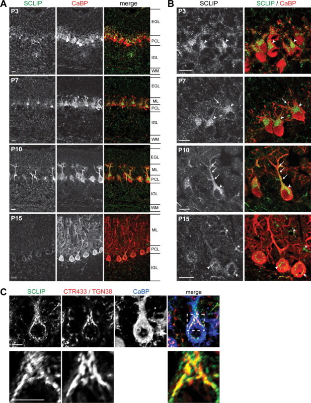
SCLIP is highly expressed in PCs and targeted to their growing dendrites during cerebellar development. A, B, Rat cerebellar slices were immunostained for SCLIP (green) and CaBP (red) at different ages of development and observed at low (A) and high (B) magnification. From P3 to P10, SCLIP is present in the different cerebellar layers but is predominantly detected in the PCL, where it appears localized in the perinuclear Golgi region (arrowheads) and developing dendrites (arrows) of PCs. At P10, SCLIP is more particularly accumulated in the principal trunk and secondary branches of PC dendritic arbors (arrows) in addition to the perinuclear Golgi region (arrowhead). At P15, however, SCLIP labeling is strongly reduced and only weakly detectable in PC somas (arrowheads). The asterisks at P15 indicate interneurons in the molecular layer also displaying SCLIP labeling in their soma [supplemental Fig. 1, available at www.jneurosci.org as supplemental material]. WM, White matter. Scale bar, 20 μm (confocal microscopy). C, P10 rat cerebellar slices were immunostained for SCLIP (green), CTR433 and TGN38 (specific markers for median- and trans-Golgi; red), and CaBP (blue). SCLIP appears in the soma as a punctuate labeling in part at the CTR433- and TGN38-labeled Golgi complex, as shown in more detail at higher magnification (bottom). Scale bar, 10 μm (confocal microscopy; z-stack projection).
