
Figure 3.
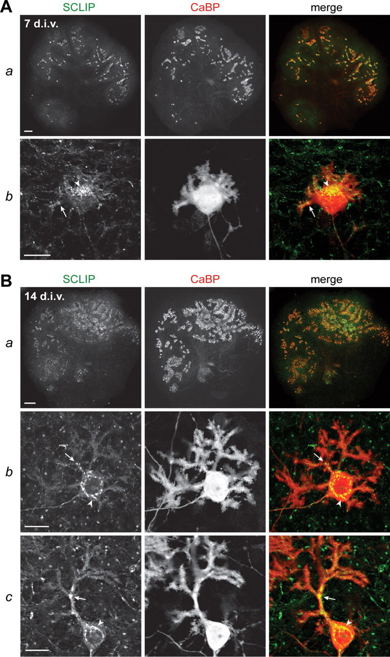
SCLIP is highly expressed in PCs in cerebellar organotypic cultures. Cerebellar organotypic cultures were immunostained for SCLIP (green) and CaBP (red) after 7 (A) and 14 (B) div. As can be seen at low magnification (Aa, Ba), SCLIP labeling follows the general pattern of PCs at both 7 and 14 div, indicating that PCs express most SCLIP in cerebellar slices cultured in vitro. Higher magnifications (Ab; Bb,c) further show SCLIP immunoreactivity in the perinuclear Golgi region (arrowheads) and dendritic processes (arrows) of PCs at the various typical developmental stages, from stage 2 to stage 4 (Fig. 5 A). Scale bars: 200 μm (lower magnifications); 20 μm (higher magnifications; confocal microscopy).