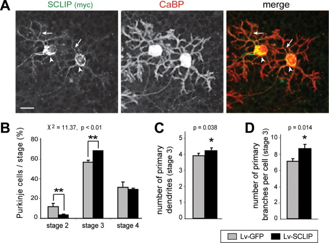
Figure 8.
SCLIP overexpression promotes PC dendritic development at stage 3 at 14 div. A, Lentiviral-mediated SCLIP-myc transduction allows SCLIP overexpression in PCs. Cerebellar organotypic cultures were infected with the Lv-SCLIP vector just after plating and immunostained for myc (green) and CaBP (red) 14 d later. A strong labeling corresponding to SCLIP-myc is detected as expected in the perinuclear Golgi region (arrowheads) and dendritic processes (arrows) of transduced PCs. Scale bar, 20 μm. B, Quantified results of three combined independent experiments illustrating the effect of SCLIP overexpression on PC developmental stages at 14 div. A higher proportion of PCs in stage 3 is observed after infection with Lv-SCLIP compared with Lv-GFP. Concomitantly, the proportion of PCs in stage 2 is similarly decreased. Shown are mean values ± SEM. Differences in the proportions of PCs in the various stages are statistically significant (**p < 0.01; χ2 independence test). C, D, Quantified results of three combined independent experiments illustrating the effect of SCLIP overexpression on PC dendrite morphology at stage 3. PCs transduced with Lv-SCLIP vector display an increased number of primary dendrites emerging from the soma (C) as well as an increased number of primary dendritic branches (D). Values shown are mean ± SEM (*p < 0.05; Mann–Whitney test).

