
Figure 8.
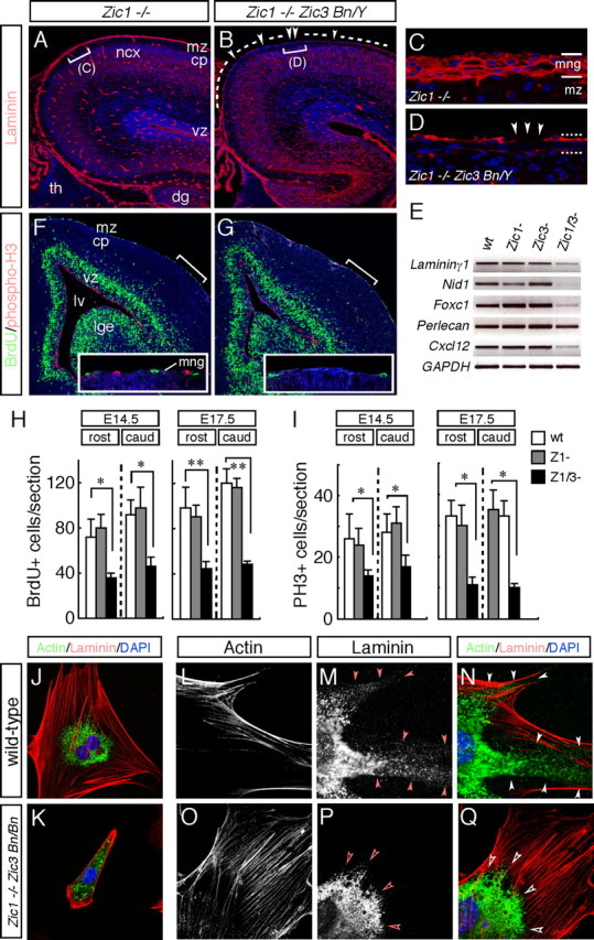
The pial BM is defective in the Zic1/3 double-mutant neocortex. A–D, Immunofluorescence detection of laminin (red) in coronal sections of the brain from the wild-type (A, C) and Zic1/3 mutant (B, D) mice at E16.5. The sections are counterstained with DAPI (blue). Control Zic1−/− show a smooth layer of basal lamina-associated laminin at the surface of the brain, as well as a basal lamina surrounding the blood vessels. In contrast, in the Zic1/3 mutant, the laminin layer at the surface of the brain is thinner compared with that in the Zic1−/− sections. C and D are high-magnification views of the indicated areas in A and B, respectively. E, Analysis for transcripts of the meningeal BM components and secreted factor. RT-PCR for Laminin γ1, Nid-1, Foxc1, Perlecan, and Cxcl12; GAPDH served as the internal control. Analysis was performed on cDNAs prepared from the meninges of wild-type (n = 6), Zic1−/− (n = 3), Zic3 Bn/Y (n = 4), and Zic1/3 (Zic1−/− Zic3 Bn/Y) (n = 3) mutants at E15.5. F, G, Decreased numbers of BrdU- and PH3-labeled proliferating meningeal cells in the Zic1/3 double-mutant mice. Proliferating meningeal cells in the S-phase were pulse labeled with BrdU for 1 h at E15.5. Immunohistochemical analysis showed decreased numbers of BrdU-labeled (green) and PH3-labeled (red) cells in the meningeal layer in the Zic1/3 mutants compared with those in the meningeal layer of the wild-type mice. The sections were counterstained with DAPI (blue). Comparable rostral areas in the Zic1/3 mutant and wild-type brains are shown. Insets are high-magnification views of the indicated areas. H, I, Quantification of the number of BrdU-labeled (H) and PH3-labeled (I) cells in the meningeal layers. BrdU- and PH3-labeled cells in the rostral and caudal regions were counted in the wild-type, Zic1−/−, and Zic1/3 mutant mice at E14.5 and E17.5. BrdU- and PH3-labeled meningeal cells were significantly decreased in the Zic1/3 mutants compared with those in the wild-type and Zic1−/− mice. The results are shown as the mean number of multiple comparable sections with SDs. *p < 0.05; **p < 0.01 by t test. J–Q, Primary meningeal fibroblasts from E14.5 wild-type (J, L–N) and Zic1/3 mutants (K, O–Q) were double stained with a laminin (J, K, M, N, P, Q) and F-actin (J, K, L, N, O, Q). N and Q are merged images of L, M and O, P, respectively. Meningeal fibroblasts were cultured for 8 h (J, K) and 3 d (L–Q). Fibroblasts from the Zic1/3 mutants spread less well and showed less proliferative activity (K). Fibrillar and punctate laminin is observed at the periphery in the wild-type meningeal fibroblasts (arrowheads in M, N), whereas it is absent from the meningeal fibroblasts of the Zic1/3 mutants (open arrowheads in P, Q). cp, Cortical plate; dg, dentate gyrus; lge, lateral ganglionic eminence; lv, lateral ventricle; mng, meninges; mz, marginal zone of the cerebral cortex; ncx, neocortex; th, thalamus; vz, ventricular zone; wt, wild type.