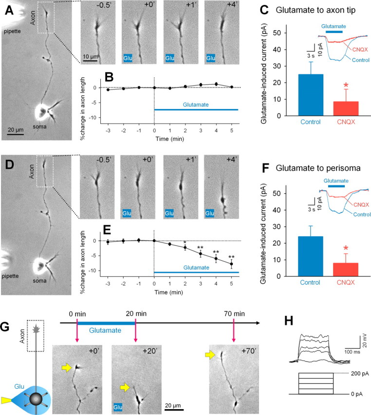
Figure 1.

Local application of glutamate to the perisomatic region induces axon retraction. A, Representative time-lapse images of the axon terminal in the inset box of the left whole-cell image. Numbers represent time (in minutes) after the start of local application of glutamate (1 mm in pipette) to the axon terminal. B, Glutamate-induced changes in axon length are summarized as means ± SEM of 12 axons. D, Representative time-lapse images of the axon terminal after local application of glutamate to the perisomatic region. The movie was taken from the same neuron as shown in the A. E, Data were summarized as means ± SEM of 10 axons. *p < 0.05, **p < 0.01 versus 0 min, paired t test. C, F, Voltage-clamp recordings revealed that local glutamate application to the axon terminal (C) and the perisoma (F) induced a CNQX-sensitive inward current. *p < 0.05 versus control, Student's t test; n = 4. The insets indicate typical traces of glutamate-evoked currents in the presence and absence of CNQX at a bath concentration of 20 μm. V H = −30 mV. G, Axons that retracted in response to perisomatic glutamate reextended when glutamate application was stopped. H, Immature granule neurons did not fire action potentials in responses to depolarizing current injection.
