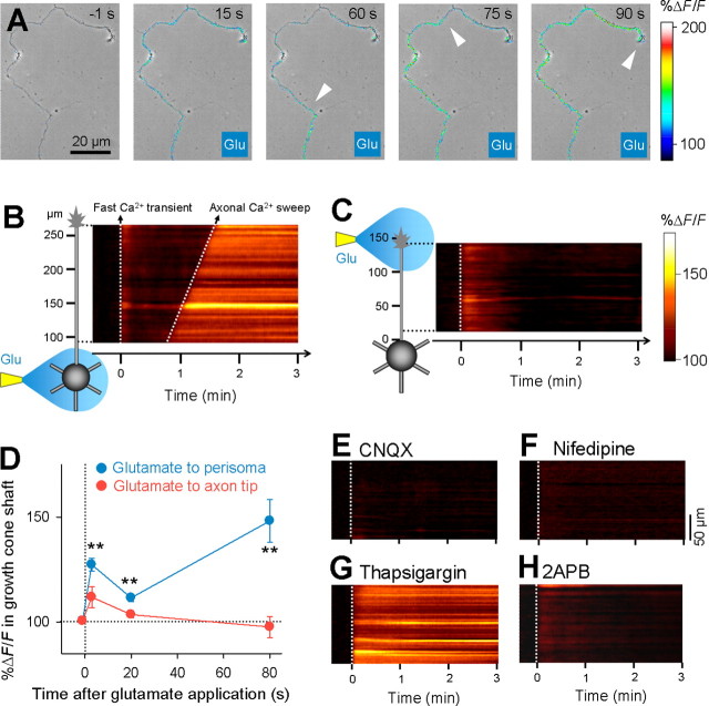
Figure 3.
Perisomatic glutamate application initiates a “wave”-like calcium signal propagating from the soma to the axon terminal. A, Time-lapse %ΔF/F confocal images of the axon of a granule cell loaded with Oregon Green 488 BAPTA-1AM are superimposed onto the same field of a phase-contrast image. Glutamate application to the perisomatic region was started at 0 s. The white arrows indicate the wave front of axonal calcium sweep. For time-lapse movie, see supplemental movie 1 (available at www.jneurosci.org as supplemental material). B, %ΔF/F of the same data as A is shown as a curvilinear line scanning along the axon. Glutamate induced two phasic calcium increases (i.e., a fast calcium transient in the entire cell and a delayed gradual calcium sweep from the soma to the axon terminal). After calcium sweep, the level of calcium was maintained for minutes. C, Local glutamate application to the axon terminal elicited only a fast calcium transient. D, Changes in axonal fluorescence intensity 20 μm from the axon terminal −1, 3, 20, and 80 s after glutamate application to the perisomatic region (blue) (n = 7) or axon tip (red) (n = 6). **p < 0.01 versus −1 s, paired t test. Error bars indicate SEM. E–H, Axonal calcium sweep was inhibited by bath application of 20 μm CNQX (E), 10 μm nifedipine (F), 1 μm thapsigargin (G), and 100 μm 2APB (H). Representative data are shown in each case, and similar results were obtained in all cases (n = 7 for CNQX; n = 7 for nifedipine; n = 7 for thapsigargin; n = 6 for 2APB).

