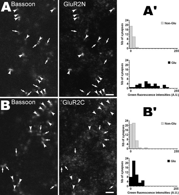
Figure 3.

Effects of epitope location on AMPA receptor detection after antigen retrieval by microwaves (P0 rat NTS). A, B, Bassoon and GluR2 channels from two triple-labeled single confocal sections (VGLUT channels not shown). GluR2 detection was performed with an antibody recognizing the extracellular N-terminal part of the protein (GluR2N) in A and with an antibody directed against the intracellular C-terminal tail (GluR2C) in B. Arrowheads and arrows indicate glutamatergic and nonglutamatergic synapses, respectively (i.e., bassoon spots with or without VGLUT immunolabeling). Note that GluR2 immunofluorescence is mainly found at glutamatergic synapses in A but not in B. A′, B′, Distributions of fluorescence intensities produced by the GluR2N antibody (A′) and the GluR2C antibody (B′) in glutamatergic and nonglutamatergic synapses. Quantification was performed as described for Figure 2A. Significant difference between the two distributions was found in A′ (p < 0.02) but not in B′ (p > 0.99). Scale bars, 2 μm. A.U., Arbitrary units.
