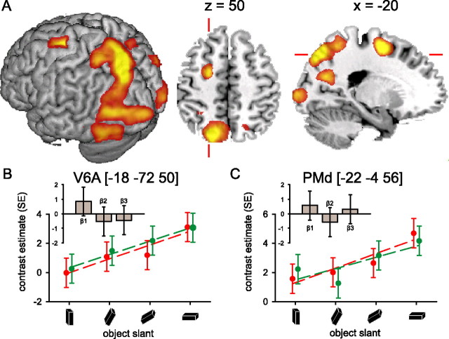
Figure 4.
Cerebral effects: object slant. A, Cerebral activity related to the prehension task increased as a function of increasing slant of the target object in occipital cortex (bilaterally), along the occipito-parietal fissure and in the anterior part of the dorsal precentral gyrus (Table 2). The image shows the relevant SPM{t} (p < 0.05 false-discovery rate corrected) superimposed on a rendered brain surface, an axial and a sagittal section. B, C, Cerebral responses over the occipito-parietal fissure (V6A) and the anterior part of the superior precentral gyrus (PMd), respectively. The graphs show parameter estimates (in SE units) for the effect evoked by the prehension task over object orientations for both viewing conditions separately. In these regions, the cerebral effects of the monocular (red) and binocular (green) viewing condition were comparable, but a strong effect of increasing object slant on cerebral activity can be seen in V6A and PMd, independent of viewing conditions. The histograms (insets) show the parameter estimates of the three basis functions used in the fMRI analysis [i.e., a canonical hemodynamic response function (β1), the weighted temporal derivative (β2), and the weighted dispersion derivative (β3)] at the corresponding local maxima. These histograms illustrate the differential BOLD amplitude (β1), latency (β2), and duration (β3) relative to the VISION by ORIENTATION interaction (i.e., the differential cerebral activity between monocular and binocular viewing conditions, increasing with object slant). The histograms show that there is no significant difference in the effect of increasing object slant between monocular and binocular viewing conditions in V6A and PMd, neither in BOLD amplitude (β1), latency (β2), nor duration (β3). Other conventions are as in Figure 3.

