Figure 5.
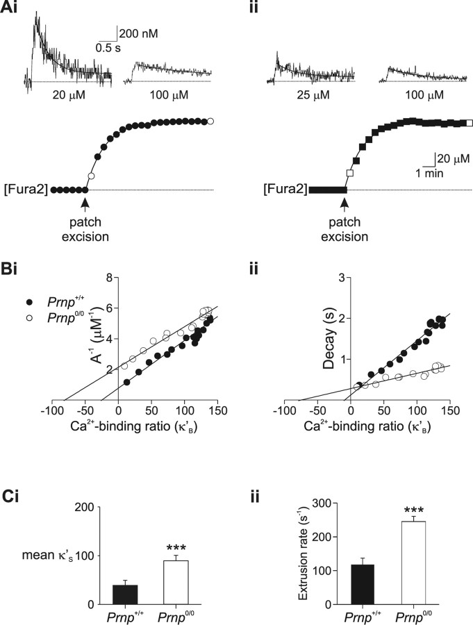
Endogenous Ca2+ buffering is enhanced in Prnp0/0 neurons. Calcium buffering of CA1 neurons was assessed by monitoring calcium changes during Fura-2 loading. Ai, The soma of a Prnp +/+ CA1 pyramidal neuron was loaded with 100 μm Fura-2 (bottom). Measurements of depolarization-evoked somatic [Ca2+]i transients were made every 30 s during loading (filled circles). [Ca2+]i transients measured directly after patch excision and after full loading (open circles) are shown in the top. The Ca2+ transients decayed with time courses of 0.37 and 1.82 s, respectively. Aii, Ca2+ buffering analysis for a Prnp0/0 neuron during loading with 100 μm Fura2 (bottom). The Ca2+ transients measured immediately after patch excision and after full loading (top, open square), decayed with time courses of 0.316 and 0.828 s, respectively. Bi, Inverse amplitude of Ca2+ changes plotted as a function of Fura-2 Ca2+ binding ratio (κ'B) for individual cells. Negative x-axis intercepts were 25 (Prnp +/+; •) and 81 (Prnp0/0; ○). Bii, Decay time constant (τ) plotted as a function of Fura-2 Ca2+ binding ratio (κ'B). Negative x-axis intercepts were 10 (Prnp +/+) and 75 (Prnp0/0), which is a measure of the ability of the neuron to buffer free Ca2+. Ci, Plot of mean endogenous binding Ca2+ binding ratio (κS) derived from inverse amplitude illustrating improved Ca2+ buffering in Prnp0/0 neurons (means of 8 Prnp +/+ and 8 Prnp0/0 cells). Cii, Plot of mean Ca2+ extrusion rate (γ) obtained from Equation 4 showing increased Ca2+ extrusion in Prnp0/0 neurons. ***p < 0.01.

