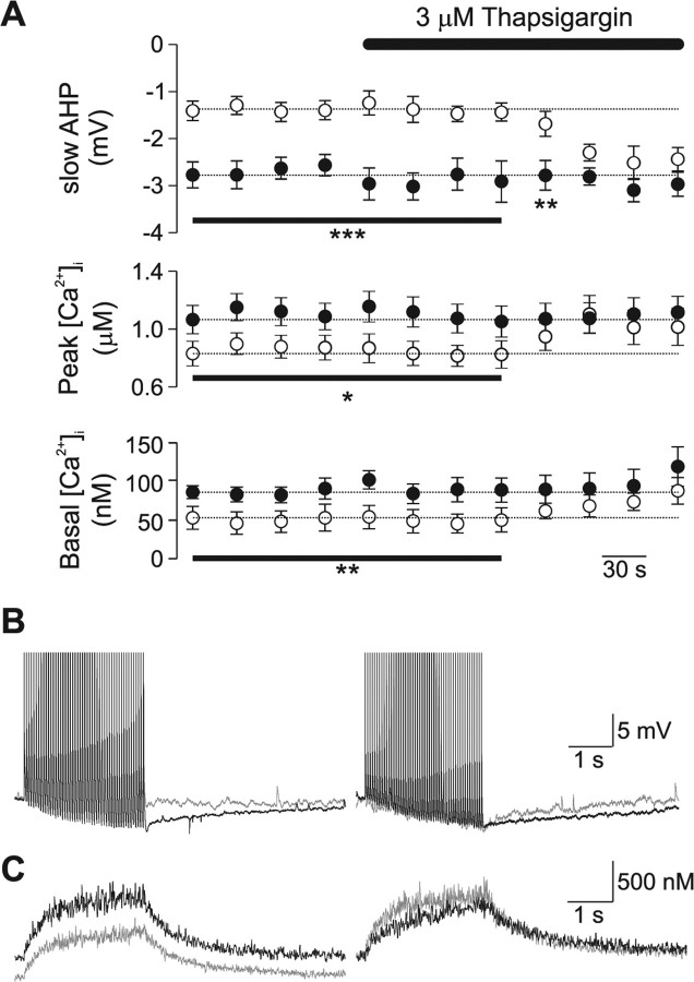
Figure 6.
Block of SERCA by thapsigargin abolishes the observed changes in slow AHP and Ca2+ signaling Prnp0/0 neurons. A, Bath application of thapsigargin (3 μm) increased slow AHP amplitude (top), peak Ca2+ levels (middle), and basal Ca2+ levels in Prnp0/0 neurons (open circles), but not Prnp +/+ neurons (filled circles). B, C, Representative traces of voltage response (B) and Ca2+ changes (C) evoked by 50 action potentials at 20 Hz; control responses are shown in the left panel, thapsigargin treated responses are illustrated in the right panel (Prnp0/0 in gray; Prnp +/+ in black). *p < 0.05; **p < 0.01; ***p < 0.005.

