
Figure 4.
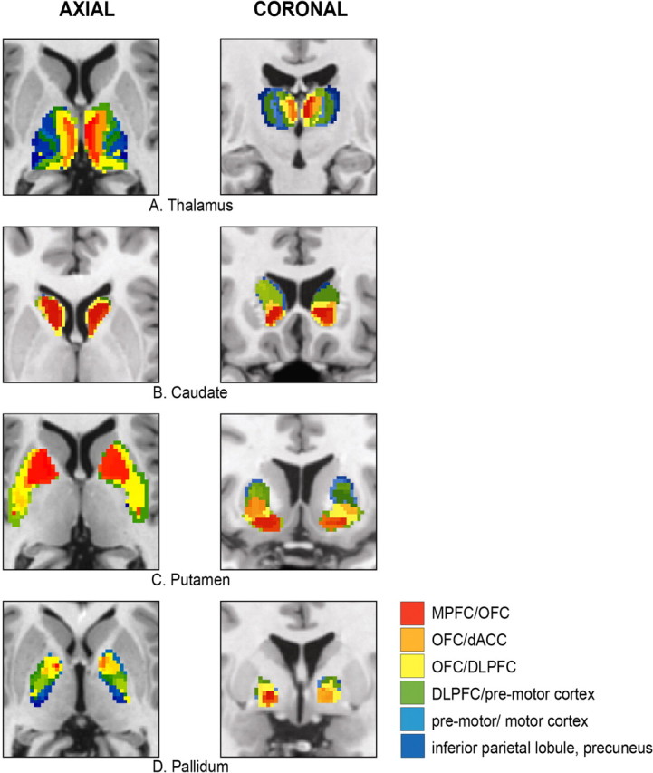
Cortical connectivity pattern of the following: A, thalamus; B, caudate; C, putamen; D, pallidum (left column, axial view; right column, coronal view). The color labeling description of the voxel connectivity profile is restricted to the larger VCPs clusters. dACC, Anterior cingulate.