
Figure 4.
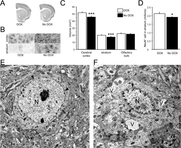
Neuropathology in the forebrain of CamDAT mice. A, C, Four weeks after DOX withdrawal, the volumes of cerebral cortex and striatum decreased by 15 and 10% respectively, whereas volume of olfactory bulb was unchanged (n = 4 per group, unpaired t test, p < 0.001 for both striatum and cortex). B, Astrogliosis was detected by GFAP staining in the cerebral cortex and striatum. n = 6 per group. D, NeuN-positive cells in the striatum decreased by 10% (n = 4, unpaired t test, p = 0.04). E, Transmission electron microscopic image of a normal striatal neuron from a CamDAT mouse under DOX treatment. F, Abnormal morphologies were found in striatum of CamDAT mice after 4 weeks of DOX withdrawal. A severely affected neuron displayed vacuolization (V) and disintegration of the nucleus (N). Arrowheads indicate the nuclear envelope. Scale bar, 2 μm.