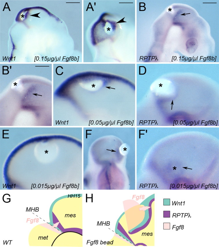
Figure 2.

Fgf8-induced upregulation of Wnt1 and RPTPλ mimics their normal spatial relationship at the MHB. A′–F′, Expression of Wnt1 (A, A′, C, E) and RPTPλ (B, B′, D, F, F′) after implantation of beads soaked in different concentrations of recombinant Fgf8b. Wnt1 expression (A, A′) is ectopically upregulated in the mesencephalon closely around a partial bead soaked in 0.15 mg/ml Fgf8b, RPTPλ expression only at a distance to the bead (B, B′). A′ is a back view of the embryo shown in A. C, D, Ectopic expression surrounding beads soaked in 0.05 mg/ml Fgf8b. E, F, F′, Expression surrounding beads soaked in 0.015 mg/ml Fgf8b. In F′, the Fgf8b-releasing bead was removed for better visual clarity. G, Schematic drawing of the gene expression patterns of Fgf8, Wnt1, and RPTPλ at the MHB of a normal chick embryo (HH15). H, Schematic drawing of Wnt1 and RPTPλ expression around a Fgf8-releasing bead fragment (pink) implanted into the mesencephalic alar plate, which acts as ectopic Fgf8 source in the dorsal midbrain. The asterisks in all panels indicate the location of Fgf8b-releasing beads. Scale bar, 100 μm.
