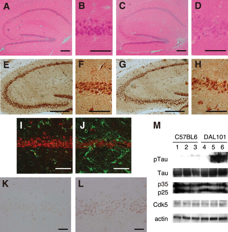
Figure 4.

Hippocampal degeneration in DAL mice. A, B, K, Twelve-month-old C57BL/6. E, F, I, Eighteen-month-old C57BL/6. C, D, L, Twelve-month-old DAL101. G, H, J, Eighteen-month-old DAL101. A, C, Hippocampus was stained with H&E. Scale bar, 200 μm. B, D, The CA1 region stained with H&E was expanded. Scale bar, 50 μm. E, G, Hippocampus was stained with the antibody to NeuN. Scale bar, 250 μm. F, H, The CA1 region stained with the antibody to NeuN was expanded. Scale bar, 50 μm. I, J, The CA1 region was stained with antibodies to GFAP (green) and NeuN (red). Scale bar, 50 μm. K, L, The CA1 region was stained with anti-phosphorylated tau antibody (AT8). Scale bar, 50 μm. M, Representative immunoblots of hippocampal lysates from 18-month-old C57BL/6 (lanes 1–3) and DAL101 (lanes 4–6) mice probed with antibodies to phosphorylated tau (pTau), pan-tau (Tau), Cdk5 activator proteins (p25 and p35), Cdk5, and actin.
