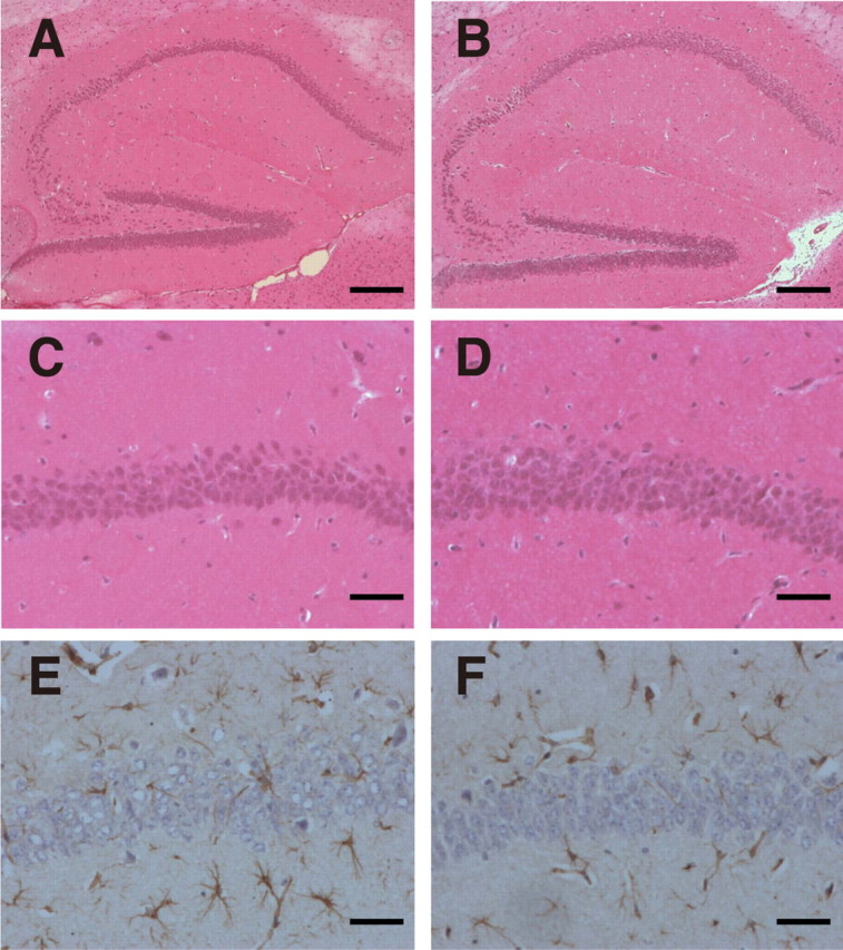
Figure 5.

No obvious hippocampal degeneration was observed in the hippocampus in young DAL mice. A, B, Hippocampus was stained with H&E. Scale bar, 200 μm. C, D, The CA1 region stained with H&E was expanded. Scale bar, 50 μm. E, F, The CA1 region was stained with anti-GFAP antibody. Scale bar, 50 μm. A, C, E, Six-month-old C57BL/6. B, D, F, Six-month-old DAL101.
