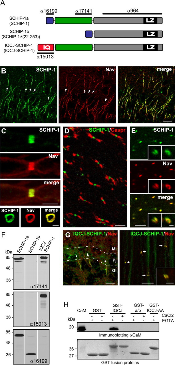
Figure 1.

IQCJ-SCHIP-1 is concentrated at AISs and NRs. A, Schematic representation of SCHIP-1 isoforms resulting from alternative splicing in mouse and human (names in human between parentheses). The three isoforms share a common C-terminal region (gray boxes), including a leucine zipper domain (black boxes; LZ). The N termini of SCHIP-1a and SCHIP-1b (blue boxes) are identical and differ from that of IQCJ-SCHIP-1, which includes an IQ motif (red box; IQ). IQCJ-SCHIP-1 and SCHIP-1a present an additional common central region (green boxes). The positions of the epitopes recognized by antibodies α17141, α15013, α16199, and α964 are indicated. B, Sections of adult mouse brain double stained for SCHIP-1 (α17141; green) and panNav (red). SCHIP-1 colocalizes with Nav at AISs (arrows) of hippocampal neurons. C, Sections of adult mouse sciatic nerves double stained for SCHIP-1 (α17141; green) and panNav (red). Top, SCHIP-1 colocalizes with Nav at NRs on longitudinal sections. Bottom, SCHIP-1 localization is restricted to the periphery of the axon and identical to Nav localization on transverse sections. D, E, Longitudinal sections of adult mouse optic nerves double stained for SCHIP-1 (α17141; green) and paranodin/Caspr (D; red) or panNav (E; red). SCHIP-1 is localized at every NR identified by paranodin/Caspr (D) or Nav staining (E). E, Insets, Higher magnification of single NR (left inset, longitudinal section; right inset, transversal section). F, Specificity of α17141, α15013, and α16199 antibodies tested by immunoblotting on lysates of COS-7 cells overexpressing one of the three mouse isoforms of SCHIP-1, as indicated. G, Double immunolabeling of adult mouse cerebellum and sciatic nerve using antibodies to IQCJ-SCHIP-1 (α15013; green) and panNav (red). IQCJ-SCHIP-1 is detectable at AISs and NRs (arrows). Inset, Higher magnification. H, Ability of IQCJ-SCHIP-1 to interact with calmodulin (CaM), as determined by GST pull-down experiments. In absence of Ca2+, CaM is immunoprecipitated by a GST fusion protein expressing the N-terminal region IQCJ-SCHIP-1 (GST-IQCJ), but not by a GST fusion protein in which the IQ residues are mutated to alanine (GST-IQCJ-AA), or by a GST fusion protein expressing the N-terminal region of SCHIP-1a/b (GST-a/b). An aliquot (250 ng) of CaM was loaded on the same gel. Scale bars: B, G, 10 μm; C (top), D, E, G (inset), 5 μm; C (bottom), E (insets), 1 μm. Gl, Granular layer; Pj, Purkinje cell layer; Ml, molecular layer.
