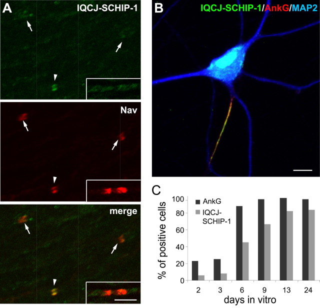
Figure 2.
IQCJ-SCHIP-1 accumulation at NRs and AISs occurs after Nav and AnkG clustering. A, Double immunolabeling of rat sciatic nerve at P0 using antibodies α17141 (green) and panNav (red). IQCJ-SCHIP-1 is detectable in a few NRs (arrowheads) but not in the large majority of them (arrows). IQCJ-SCHIP-1 is also absent from most heminodes (insets). B, Cultured hippocampal neurons at DIV 23 stained with α17141 (green) and antibodies to AnkG (red) and MAP2 (blue). IQCJ-SCHIP-1 is enriched at the AISs identified by AnkG staining. C, Percentage of neurons with AISs immunopositive for IQCJ-SCHIP-1 or AnkG during in vitro maturation of hippocampal neurons. Results are expressed as a percentage of the total number of cells (n = 800–2000 cells per time point in 2 independent experiments). Scale bars: A, 5 μm; B, 10 μm.

