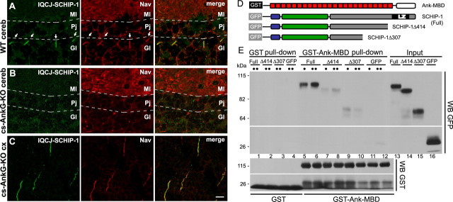
Figure 3.
IQCJ-SCHIP-1 localization at AISs of Purkinje cells is lost in cerebellum-specific AnkG knock-out mice. A–C, Brain sections of wild-type (A; WT) and cerebellum-specific AnkG knock-out (B, C; cs-AnkG-KO) mice double stained with α17141 (green) and panNav (red). A, B, IQCJ-SCHIP-1 and Nav are not detectable at AISs of Purkinje cells of mutant mice (B), in contrast with wild-type mice (A, arrows). C, IQCJ-SCHIP-1 and Nav labeling is normal at AISs of cortical neurons in cs-AnkG-KO mice. D, Constructions used for GST pull-down experiments were as follows: GST-Ank-MBD includes the conserved MBD of ankyrins (red boxes). GFP-fusion proteins encompass SCHIP-1a (full) and SCHIP-1aΔ414 and SCHIP-1aΔ307 (deleted of the 73 and 180 C-terminal residues, respectively). E, GST-Ank-MBD (lanes 5–12), but not GST alone (lanes 1–4), precipitated GFP-SCHIP-1 (full; lanes 5–6) but not the truncated forms (lanes 7–10), nor GFP alone (lanes 11–12). For each fusion protein, 1:12 (•) or 1:6 (••) of the lysate was loaded. Inputs (2%) were loaded on the same gel (lanes 13–16). Scale bar, 10 μm. cereb, Cerebellum; cx, cortex; Gl, Granular layer; Pj, Purkinje cell layer; Ml, molecular layer.

