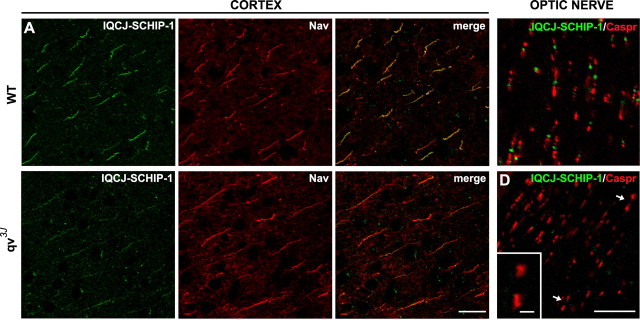
Figure 4.
Clustering of IQCJ-SCHIP-1 at NRs and AISs is disrupted in qv3J mice. A, B, Sections of brains of wild-type (A; WT) and qv3J (B; qv3J) mice double stained with α17141 (green) and panNav (red). IQCJ-SCHIP-1 staining at AISs of cortical neurons is dramatically reduced in 2-month-old qv3J mice (B) compared with wild-type mice (A), whereas Nav staining was unchanged. Quantification of immunofluorescence (t test) was as follows: IQCJ-SCHIP-1, WT = 100.5 ± 5.5 arbitrary units (a.u.), qv3J = 50.1 ± 3.5 a.u., p < 0.001; Nav, WT = 131.5 ± 3.6 a.u., qv3J = 146.3 ± 6.6 a.u., p > 0.05, n = 20 AISs per phenotype. IQCJ-SCHIP-1 staining was completely lost at AISs of 6-month-old qv3J mice (data not shown). C, D, Longitudinal sections of optic nerves of wild-type (C; WT) and qv3J (D; qv3J) mice double stained with α17141 (green) and paranodin/Caspr (red). IQCJ-SCHIP-1 is not detectable at NRs identified by paranodin/Caspr labeling in qv3J mice (D; arrows), whereas it is clearly detectable at NRs of wild-type mice (C). D, Inset, Higher magnification of NRs lacking IQCJ-SCHIP-1 staining. Scale bars: A, B, 20 μm; C, D, 10 μm; D, Inset, 2 μm.

