
Figure 1.
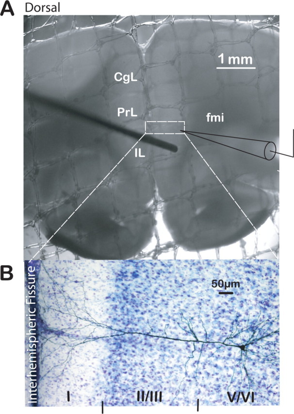
Brain slice preparation of the rat PFC used to study putative BLA–mPFC synapse. A, Photograph of a PFC slice submerged in our recording chamber depicting the position of stimulating (from the left) and recording (from the right) electrodes for EPSC recordings. CgL, Cingulate cortex; fmi, forceps minor. Scale bar, 1 mm. Dashed rectangle depicts area expanded below of typical biocytin-filled, layer V, PrL pyramidal neuron from which recordings were made. B, Photograph of DAB-stained layer V pyramidal neuron filled with biocytin in the cresyl violet-counterstained mPFC. Cortical layers are indicated by roman numerals at the bottom of the photograph. Scale bar, 50 μm.