Figure 4.
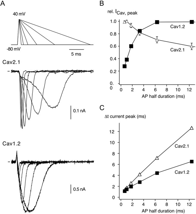
Current responses of Cav2.1 and Cav1.2 channels to AP stimuli. A, Representative current traces evoked in Cav2.1 (middle) and Cav1.2 (bottom) channels by AP-like waveforms (top) comprising a 0.52 ms depolarizing ramp from −80 to 40 mV and a repolarizing ramp from 40 to −80 mV with variable duration of 0.76, 1.52, 3.04, 6.08, 12.16, or 24.32 ms; these durations correspond to AP half durations of 0.64, 1.02, 1.78, 3.3, 6.34, and 12.42 ms. Note the distinct current profiles generated by the two Cav subtypes. B, Peak Ca2+ currents elicited by AP-like waveforms normalized to the maximal current amplitude obtained with the AP series indicated in A. Data points are the mean ± SEM of six experiments for each Cav channel. C, Period between peak current and start of the AP command (Δt peak current) as a function of the AP half duration. Data points are mean ± SEM of six experiments for each Cav channel. Note the distinct increase in Δt peak current for both Cav subtypes.

