
Figure 5.
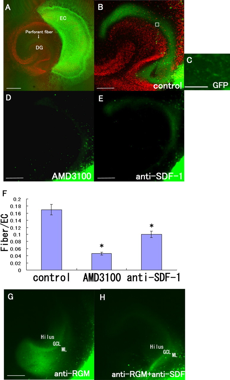
SDF-1α is necessary for the projection of perforant fibers from the EC to the DG. A, The GFP-labeled EC (green) and DG (red; stained with anti-Prox1 antibody) slices were obtained from rats at postnatal days 0–1. B, D, E, The tissue slices were incubated for 9 d in the absence (B) or presence of 50 μg/ml AMD3100 (CXCR4 antagonist) (D) or 60 μg/ml anti-SDF-1 antibody (E). A, B, Perforant fibers (arrow) running from the EC to the DG are visible. C, High-magnification view of a square in B. D, E, Treatment with AMD3100 (SDF-1 receptor antagonist) or the anti-SDF-1 antibody reduced the projection of the perforant fibers from the EC to the DG. F, The fluorescence intensity of the perforant fibers in the DG was measured. The data are represented as the mean ± SEM of four samples (8 fields of 25 μm2 each) of each group. The perforant fiber projection was significantly lower in the AMD3100- and anti-SDF-1 antibody-treated groups. The asterisks (*) indicate statistical significance (p < 0.05) (one-way ANOVA followed by Scheffé's multiple-comparison test). G, H, The tissue slices were incubated with anti-RGM antibody (G) or anti-RGM antibody and anti-SDF-1 antibody (H). Note the loss of the layer-specific termination of GFP-positive fibers after treatment with the anti-RGMa antibody. GCL, Granule cell layer; ML, molecular layer. Scale bars: A, 500 μm; B, D, E, G, 200 μm; C, 12 μm.