Figure 8.

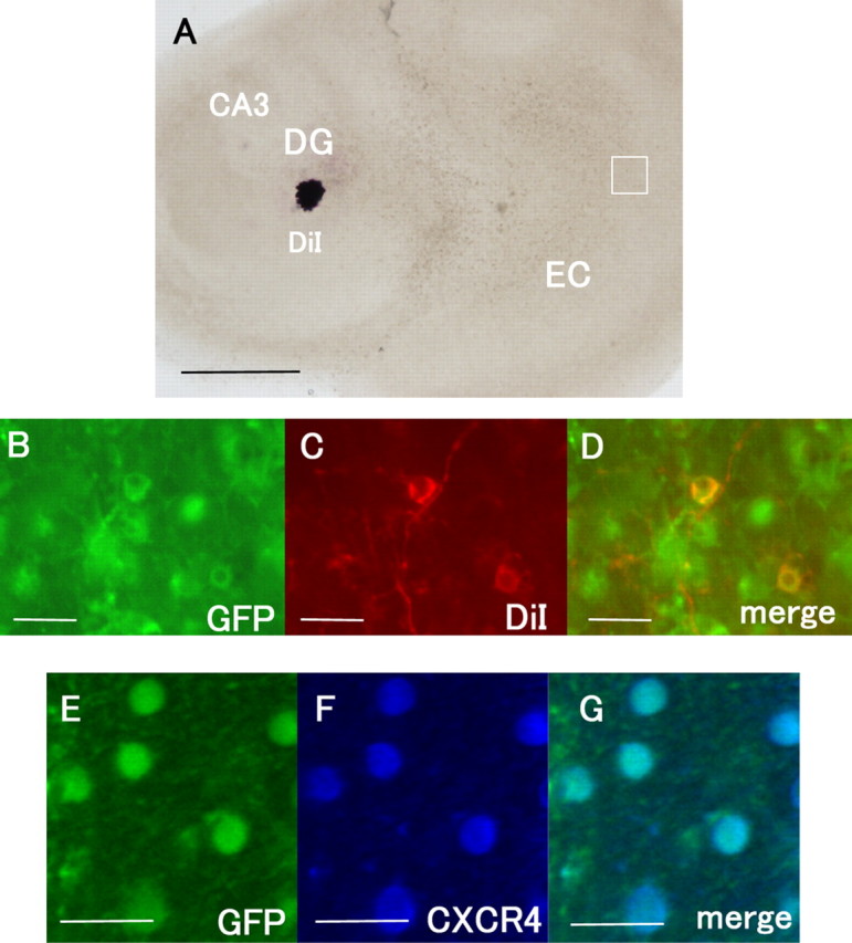
The EC neurons that project into the DG express CXCR4. A, DiI was placed into the DG to label the perforant fibers. B–D, The GFP signals in the EC were colocalized with those of DiI. E–G, The immunoreactivity for CXCR4 (blue) is observed in the GFP-positive cells in the EC. Scale bars: A, 500 μm; B–G, 20 μm.
