
Figure 1.
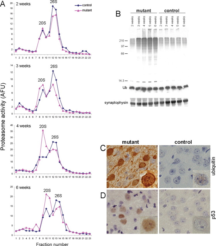
Ablation of Psmc1 causes 26S proteasome depletion. A, Representative proteasome profiles from cortex of 2-, 3-, 4-, and 6-week-old control and Psmc1 fl/fl;CaMKIIα-Cre (mutant) mice after glycerol density gradient centrifugation. Fractions 1 and 23 correspond to the top (10%) and bottom (40%) of the gradient, respectively. 20S and 26S proteasomal distribution was revealed by succinyl-Leu-Leu-Val-Tyr-7-amido-4-methylcoumarin hydrolysis activity of gradient fractions. Individual experiments at different ages are not directly comparable because of, for example, the amount of protein loaded and slight variability in fraction collection. AFU, Arbitrary fluorescent unit. B, Accumulation of polyubiquitinated proteins, but not free ubiquitin (Ub), in the Psmc1 fl/fl;CaMKIIα-Cre (mutant) cortex with increasing age, shown by Western analysis of cortex homogenates with an anti-ubiquitin antibody. Anti-synaptophysin was used as a loading control. C, D, Immunohistological staining of cortex sections from 6-week-old control and Psmc1 fl/fl;CaMKIIα-Cre (mutant) mice using anti-ubiquitin (C) and anti-p53 (D) antibodies (40×). Higher-magnification images are inset into the representative sections.