
Figure 5.
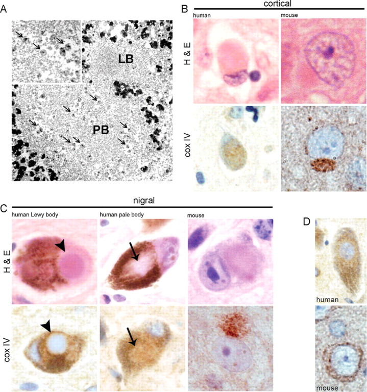
Mitochondria in human and mouse Lewy bodies. A, EM showing mitochondria (arrows) among filamentous material in a human nigral PB, which is adjacent to a classical LB that is devoid of mitochondria. Enlarged view of mitochondria within the pale body is shown in the inset. B, C, Shown is H&E and cox IV immunostaining of cortical Lewy bodies (B) and nigral Lewy and pale bodies (C) from human disease and Psmc1 fl/fl;CaMKIIα-Cre and Psmc1 fl/fl;TH Cre mice (100×). The arrows and arrowheads identify the pale and classical dense core Lewy bodies of human disease, respectively. D, The normal pattern of cox IV in human and mouse neurons (100×). The heavy brown pigment in H&E human nigral neurons is neuromelanin.